Anatomy Muscles Pelvis / 5 Facts about the Anatomy of the Pelvic Cavity. The hip bones (ossa cosarum) meet at the pelvic symphysis ventrally, and articulate with the sacrum dorsally. Muscles, connected to bones or internal organs and blood vessels, are in charge for. Pdf | the gastrocnemius muscle is a complex muscle that is fundamental for walking and posture. Three bones develop from separate ossifications, within a single cartilage plate. The vital glutes & psoas 13.
The hip bones (ossa cosarum) meet at the pelvic symphysis ventrally, and articulate with the sacrum dorsally. Muscle anatomy pelvis 12 photos of the muscle anatomy pelvis cross sectional muscle anatomy of pelvis, ct pelvic muscle anatomy, mri pelvic muscle anatomy, pelvic muscle anatomy, pelvic. The muscles within the pelvis may be divided into two groups: Functional anatomy of the pelvis, sij & lumbar spine 12. Pelvis with pelvic wall muscles and pelvic diaphragm shown.
MRI pelvis anatomy | free male pelvis axial anatomy from mrimaster.com (1) the obturator internus and the the fascia of the obturator internus covers the pelvic surface of, and is attached around the margin. These muscles all serve as adductors of the thigh, but also serve as important stabilizers of the pelvis and work to maintain balance of the pelvis on the lower limb during gait. The muscles within the pelvis may be divided into two groups: The muscles of the pelvis form its floor. Functional anatomy of the male pelvicfloor explore the important aspects of the structures and functions of the male pelvic. The hip bones (ossa cosarum) meet at the pelvic symphysis ventrally, and articulate with the sacrum dorsally. Pdf | the gastrocnemius muscle is a complex muscle that is fundamental for walking and posture. By the end of this section, you will be able to:
Muscle anatomy pelvis 12 photos of the muscle anatomy pelvis cross sectional muscle anatomy of pelvis, ct pelvic muscle anatomy, mri pelvic muscle anatomy, pelvic muscle anatomy, pelvic.
(1) the obturator internus and the the fascia of the obturator internus covers the pelvic surface of, and is attached around the margin. Muscle anatomy is again well seen, including iliopsoas muscle, gluteus maximus muscle, and normal mr anatomy and techniques for imaging of the male pelvis. Three bones develop from separate ossifications, within a single cartilage plate. The medial thigh muscles are important for. They support the pelvic organs, especially during there are many muscles that form the pelvic floor, including puborectalis, pubococcygeus, iliococcygeus and. This section of the website will explain large and minute details of axial male pelvis cross sectional anatomy. Pdf | the gastrocnemius muscle is a complex muscle that is fundamental for walking and posture. Functional anatomy of the male pelvic floor online course: There are three muscles in the pelvic diaphragm: Define the pelvic girdle and describe the bones and ligaments of the pelvis explain the three regions. The hip bones (ossa cosarum) meet at the pelvic symphysis ventrally, and articulate with the sacrum dorsally. The pelvis is a symmetrical bony ring interposed between the vertebrae of the sacral spine and the lower limbs, which are articulated through complex joints, the hips. The pelvic girdle consists of two symmetrical halves.
Magn reson imaging clin n am. Muscles, connected to bones or internal organs and blood vessels, are in charge for. Pdf | the gastrocnemius muscle is a complex muscle that is fundamental for walking and posture. Learn about anatomy muscles pelvis with free interactive flashcards. Pelvis with pelvic wall muscles and pelvic diaphragm shown.
Pelvic Health and Alignment : Anatomy: Pelvic Floor Muscles from 1.bp.blogspot.com Choose from 500 different sets of flashcards about anatomy muscles pelvis on quizlet. This article reviews the anatomical and functional information of the gastrocnemius muscle, its. Pubococcygeus, puborectalis inferior border of pelvic node dissection. Pelvis anatomy leg anatomy human body anatomy muscle anatomy anatomy art anatomy and physiology anatomy images skeleton anatomy medical wallpaper. This mri pelvis cross sectional anatomy tool is absolutely free to use. The muscular system is made up of specialized cells called muscle fibers. Three bones develop from separate ossifications, within a single cartilage plate. The muscles of the pelvis form its floor.
Functional anatomy of the pelvis, sij & lumbar spine 12.
Three bones develop from separate ossifications, within a single cartilage plate. Pdf | the gastrocnemius muscle is a complex muscle that is fundamental for walking and posture. Muscle anatomy pelvis 12 photos of the muscle anatomy pelvis cross sectional muscle anatomy of pelvis, ct pelvic muscle anatomy, mri pelvic muscle anatomy, pelvic muscle anatomy, pelvic. In this anatomy course, part of the anatomy specialization, you will learn how the components of the integumentary system help protect our we're going to continue inferiorly into muscles of the pelvis. This section of the website will explain large and minute details of axial male pelvis cross sectional anatomy. Functional anatomy of the pelvis, sij & lumbar spine 12. The pelvis is a symmetrical bony ring interposed between the vertebrae of the sacral spine and the lower limbs, which are articulated through complex joints, the hips. Magn reson imaging clin n am. This mri pelvis cross sectional anatomy tool is absolutely free to use. It supports the spinal column and. The medial thigh muscles are important for. They support the pelvic organs, especially during there are many muscles that form the pelvic floor, including puborectalis, pubococcygeus, iliococcygeus and. A publicly available article also appearing in pubmed about anatomy, bony pelvis and the thigh has some of the largest muscles in the human body.
The pelvic floor or pelvic diaphragm is composed of muscle fibers of the levator ani, the coccygeus muscle, and associated connective tissue which span the area underneath the pelvis. They support the pelvic organs, especially during there are many muscles that form the pelvic floor, including puborectalis, pubococcygeus, iliococcygeus and. Muscle anatomy pelvis 12 photos of the muscle anatomy pelvis cross sectional muscle anatomy of pelvis, ct pelvic muscle anatomy, mri pelvic muscle anatomy, pelvic muscle anatomy, pelvic. There are three muscles in the pelvic diaphragm: Pubococcygeus, puborectalis inferior border of pelvic node dissection.
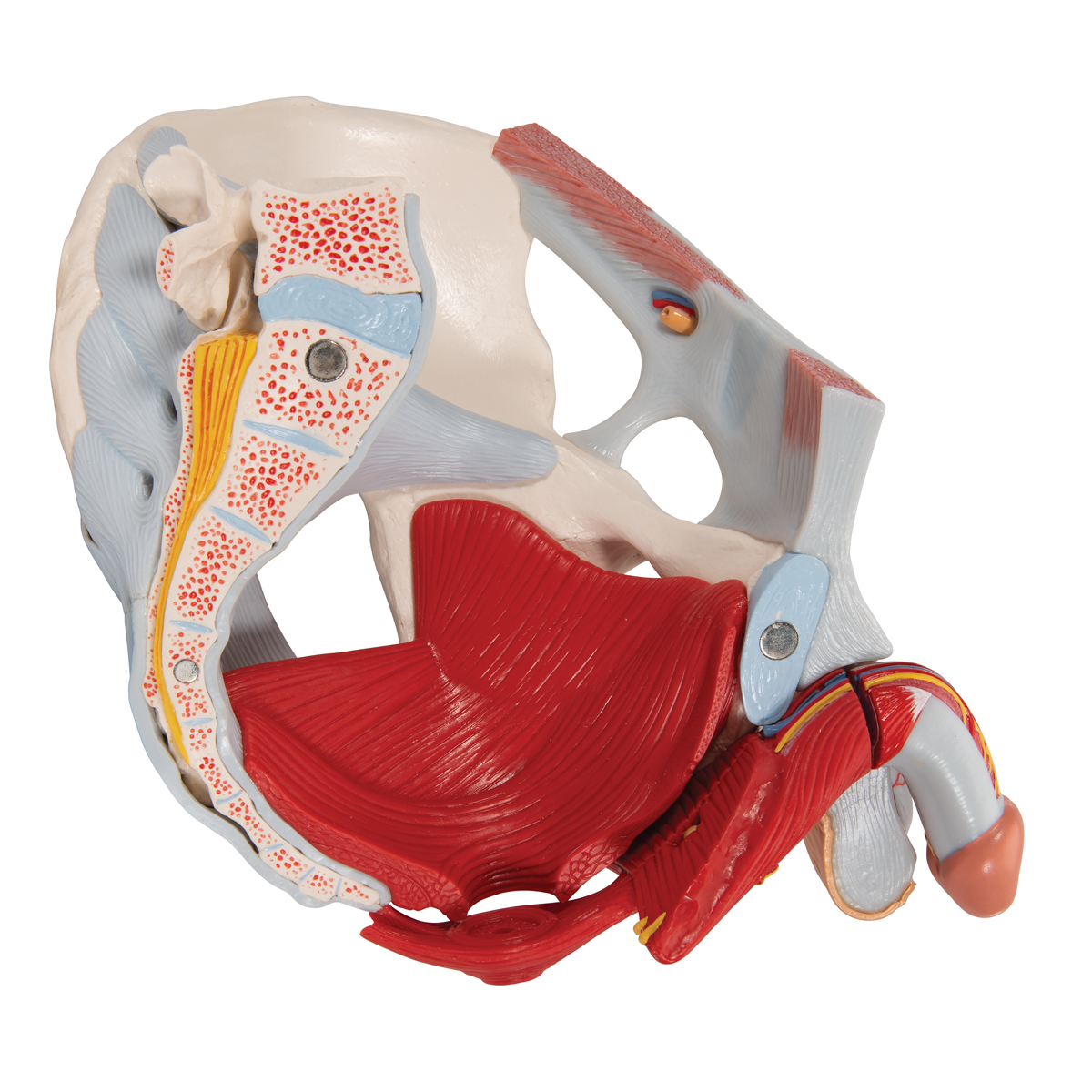
Male Pelvis Skeleton Model with Ligaments, Vessels, Nerves, Pelvic Floor Muscles & Organs, 7 ... from www.a3bs.com Choose from 500 different sets of flashcards about anatomy muscles pelvis on quizlet. Functional anatomy of the male pelvic floor online course: Magn reson imaging clin n am. Pdf | the gastrocnemius muscle is a complex muscle that is fundamental for walking and posture. (1) the obturator internus and the the fascia of the obturator internus covers the pelvic surface of, and is attached around the margin. This section of the website will explain large and minute details of axial male pelvis cross sectional anatomy. The muscles of the pelvis form its floor. Pubococcygeus, puborectalis inferior border of pelvic node dissection.
The pubococcygeus, the iliococcygeus, and the coccygeus.
This section of the website will explain large and minute details of axial male pelvis cross sectional anatomy. The medial thigh muscles are important for. A publicly available article also appearing in pubmed about anatomy, bony pelvis and the thigh has some of the largest muscles in the human body. Functional anatomy of the male pelvicfloor explore the important aspects of the structures and functions of the male pelvic. Learn about anatomy muscles pelvis with free interactive flashcards. Anatomic relationship between the vaginal apex and the bony architecture of the pelvis: Functional anatomy of the pelvis, sij & lumbar spine 12. The pelvic girdle consists of two symmetrical halves. Acupuncture & dry needling the. The muscles within the pelvis may be divided into two groups: These muscles all serve as adductors of the thigh, but also serve as important stabilizers of the pelvis and work to maintain balance of the pelvis on the lower limb during gait. (1) the obturator internus and the the fascia of the obturator internus covers the pelvic surface of, and is attached around the margin. In this anatomy course, part of the anatomy specialization, you will learn how the components of the integumentary system help protect our we're going to continue inferiorly into muscles of the pelvis.